Tag Archives: vomiting

Coughing cats

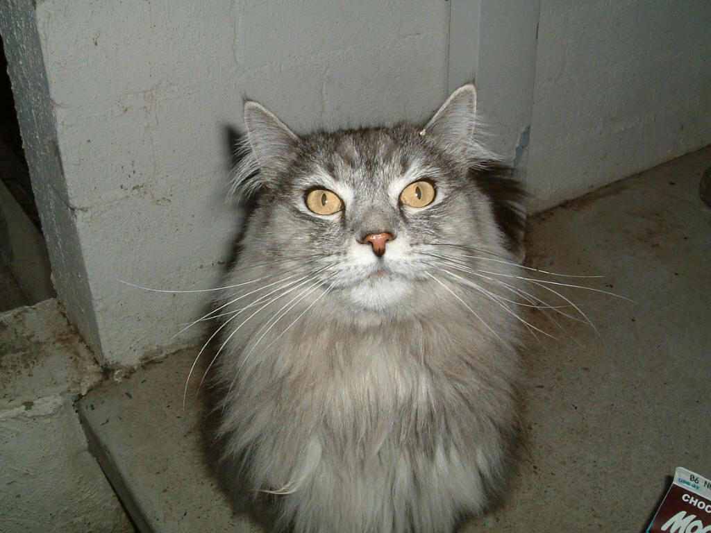
At first Gus’s carer thought he had hair balls. So did the friends she asked. He gagged and convulsed and brought up froth. She gave him some laxative paste.

Everything in the litter tray seemed normal and for a while Gus seemed OK.

When she rushed off to work he was curled up on the lounge in the sun room as usual.

But the gagging started up again, especially at night. She noticed that he wasn’t eating all his dinner and sometimes he stopped in the middle of the gagging and breathed heavily.

One night he crept on to the end of the bed and wheezed and gasped for breath until she was sure he was choking to death.

Next morning she rushed him into us. We X-rayed his chest and found a very hazy lung and signs of chronic bronchitis.

We took samples from Gus’s lungs and found he had pneumonia. Gus had developed an airway and lung infection on top of the chronic bronchitis.

Cats get asthma and bronchitis, just like humans do. For some it is worse when there are lots of pollens blowing about, for others being cooped up inside with the stagnant air and dust mites in winter set the wheezing and coughing off.

His carer remembered that he had always had a bit of a wheeze, especially in spring and early summer. She hadn’t thought much of it.

It is very easy to confuse coughing with vomiting or regurgitation. Usually food or bile will come up at some stage with vomiting. Vomiting cats often lose their appetite or have diarrhoea as well. Coughing cats don’t go off their food unless they develop an infection as well.

Some asthmatic cats have life threatening breathing difficulties if they are not treated adequately. If you notice your cat coughing, gagging, breathing with difficulty, especially with the mouth open and the neck extended, contact your vet.

Check out Fritz the Brave for reliable information and support if your cat has asthma or bronchitis in cats.

Gus is back to his irascible self after a long course of antibiotics. He’s getting used to a puffer and spacer, and quite likes all the attention we give him.

 

Bloat or the dreaded GDV

In bloat, also known as gastric dilation, a dog’s stomach is over distended with gas. Often it twists as well, sending the dog into shock. When this happens it is called gastric dilation and volvulus, or GDV.

Factors thought to increase the risk of bloat and GDV include:

  • Gulping air down with food
  • Strenuous exercise soon after a meal
  • Leanness
  • Drinking a lot of water immediately after a large meal of dry food
  • Increasing age
  • An anxious or timid temperament

Factors thought to decrease the risk of bloat and GDV include:

  • Canned food
  • Table scraps in the diet
  • Happy or easy-going temperament
  • Two or more meals per day

Male, deep-chested large breeds like Great Danes, Irish Setters, German Shepherds, Afghan Hounds, Basset Hounds and Rottweilers are most often affected.

A bloating dog becomes uncomfortable and restless immediately after a meal. The left side of the abdomen distends and the dog tries to vomit. Some dogs adopt a “praying position” because of the pain.

An x-ray confirms the bloat and tells us if the stomach has twisted.

Affected dogs go into shock because the distended stomach puts pressure on the large veins in the abdomen that carry blood back to the heart. The output of blood from the heart drops and the supply of oxygen and nutrients to the rest of the body comes to halt.

The blood supply to the stomach wall also drops and with increasing internal pressure the stomach wall begins to die and may rupture.

If the stomach twists the blood supply to the spleen is cut off and it swells and soon dies.

In many dogs the heart goes out of its normal rhythm either before, during or up to 2 days after surgery.

When the stomach is distended, digestion stops. Toxins accumulate, move into the circulation and activate chemicals which cause inflammation. Blood clots then form within blood vessels. This is called disseminated intravascular coagulation (DIC) and is usually fatal.

How can we save the dog’s life?

We must act quickly to save the dog’s life.

  1. Large quantities of intravenous fluids treat the shock.
  2. The pressure in the stomach is relieved with a tube passed from the mouth to the stomach or with a needle pushed through the skin into the stomach.
  3. The stomach is returned to its normal position in surgery.
  4. Any dead areas of the stomach are cut out. If the spleen has died it is also removed.
  5. The stomach is stitched to the abdominal wall (gastropexy) to reduce the chance of recurrence of GDV.
  6. We monitor the heart for abnormalities in its rhythm (arrhythmias) before, during and after surgery, and treat as necessary.

What is the survival rate?

The survival rate depends on the severity of the distention, the degree of shock, how quickly treatment is begun, and the presence of other diseases, especially those involving the heart. Approximately 60 to 70% of treated dogs survive.

How do we prevent bloat and GDV happening again?

Gastropexy does not prevent bloat but it is usually successful in preventing twisting of the stomach and spleen. We assess each individual’s meal time habits for any of the predisposing factors mentioned above.


Kidney failure in cats

Mittens getting the right balance

What do kidneys do?


The kidneys remove waste products from the blood stream, regulate fluid and electrolyte balance, maintain the acid-base balance of the body and remove toxins and drugs. They also help maintain blood pressure and stimulate blood cell production.

What happens when my cat’s kidneys fail?

Signs of kidney failure don’t appear until at least 70% of kidney function is lost. Chronic kidney failure is known as renal failure.

Kidney damage accumulates for years before we see any signs. Even then the early signs of kidney failure – increased thirst and urine production – are not easily recognised in our feline friends.

You may notice an increasingly wet litter tray if your cat is only indoors. However if you have other cats you may not pick up increased urine production in a single cat.

Cats often drink from multiple water sources making it difficult to recognise increased consumption.

Other signs of kidney failure such as weight loss and poor coat quality are often put down to normal ageing.

Often the first thing we see is a cat off her food, vomiting, depressed and dehydrated. The kidneys are already badly affected by this stage.

How do we diagnose kidney failure?

We diagnose and stage kidney failure with blood tests for the two waste products, urea and creatinine and a urine analysis to measure the kidneys ability to concentrate urine. We also  check the urine for protein loss or a urinary tract infection.

Tests for other substances like potassium, phosphorus and calcium as well as blood cell counts help us decide on the best course of treatment.

Could it have been diagnosed earlier?

Because signs of kidney failure and rises in blood urea and creatinine are not evident until significant loss of kidney function has occurred early diagnosis is difficult. However, we strongly recommend at least annual blood and urine tests, as well as regular body weight checks. If urine concentrating ability is deteriorating or the creatinine is trending up we are able to slow down the progression of the disease with a special kidney protective diet. Any weight loss in a cat should be fully investigated.

What treatments are available?

After initial hospitalisation to treat dehydration and electrolyte disturbances, most cats are managed with a diet change and one or two other medications.

  • Low protein and phosphorus diets lower the level of waste products in the bloodstream.  Try a few of the ready made kidney diets like Royal Canin renal or Hills k/d to find one your cat likes. Once your cat accepts the diet it must be her sole source of nutrition. Although if renal failure is advanced and your cat’s appetite is poor, any diet that the cat enjoys is acceptable.
  • If blood phosphorus levels remain above normal after a few weeks on the special diet we add phosphate binders like Ipakitine to the food. Reducing blood phosphorus makes your cat feel better and slows progression of the disease.
  • Urinary tract infections are common in kidney failure and are treated with antibiotics.
  • Cats with renal failure lose potassium in the urine leading to muscle weakness, stiffness, a poor coat and exacerbation of the kidney failure. The special kidney diets contain extra potassium but sometimes we have to add more.
  • Severely affected cats need extra fluids. We can teach you to administer      subcutaneous fluids at home to reduce dehydration in advanced cases.

FRESH WATER MUST BE AVAILABLE AT ALL TIMES

How long can I expect my cat to live?

Unfortunately aged kidneys do not recover. However, we can slow the progress of the disease and improve your cat’s well being with treatment and regular checkups. We check  phosphorous and potassium levels to see if your cat requires supplements and check for urinary tract infections at least every 3 months. This should give your cat a good stretch of high quality, active life.


Roundworm

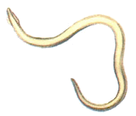
Roundworms, also known as ascarids, have round bodies and are 7-12 cm long. They live in  dogs’ intestines on partially digested food.

Pups infested with roundworms lose weight and look pot-bellied. They lose their appetites and may vomit or develop diarrhoea. Serious roundworm infestations obstruct or twist the bowel.

Most puppies acquire roundworm infection from their mothers before birth and then through the milk afterward.

Both puppies and adult dogs are also infected by swallowing roundworm eggs from bowls, hair or dirt contaminated with faeces. They can also be infected by roundworm larvae encysted in the organs of prey like mice, cockroaches, earthworms and chickens.

Ingested larvae and eggs are activated in the dog’s stomach and small intestine and migrate through the muscle, liver, and lungs. After several weeks, the larvae make their way back to the intestine to mature. When these worms begin to reproduce, new eggs will pass in the dog’s faeces, and the life cycle of the parasite is completed.

Roundworm eggs in the faeces confirm roundworm infestation. Because roundworm don’t produce many eggs we sometimes have to examine several fecal samples before we find eggs. Occasionally, we see mature worms in the dog’s vomit or faeces.

Treatment with broad-spectrum wormers like Milbemax or Drontal eliminates adult roundworm. No treatment reliably kills the migrating larvae so we give two or three treatments 2 weeks apart catch them as they mature into adult worms.

The eggs are resistant to most disinfectants and survive in quite harsh environmental conditions. They remain infective to dogs and humans for years.

Regular removal of dog faeces and deworming as detailed below prevent reinfection.

Roundworms are a risk to human health.  Children and people who are immunosuppressed or on immunosuppressive drugs are particularly vulnerable. The roundworm eggs only develop as far as larvae in people. As the larvae migrate through the body they can cause damage, particularly to the eye. Worming people does not kill the larvae as only adult worms are susceptible to wormers. To prevention human infestation

  • Deworm pups fortnightly until 12 weeks of age, then monthly until 6 months of age. Adult dogs must be dewormed every 3 months
  • Remove and dispose of dog faeces promptly
  • Teach children to wash their hands thoroughly after playing with dogs
  • Deworm pregnant bitches in late pregnancy and then every time her pups are dewormed, as above
  • Discourage dogs from defecating in children’s play areas


Parvovirus in dogs

Parvovirus causes severe vomiting and diarrhoea and is very contagious. The virus may infect a whole litter of pups from an unvaccinated bitch. Dogs less than one year old are the most vulnerable to the virus.

Dogs go off their food and start to vomit within a few days of infection. An astute owner will notice a drop in appetite, depression, and fever before the vomiting and diarrhoea start. The diarrhoea often contains blood and mucus, and many dogs suffer severe pain in the belly.

How does a dog become infected with parvovirus?

The faeces of an infected dog is high in virus. Direct contact between dogs is not required to spread the virus.

Another dog is infected by licking the virus off food dishes, hair, the ground, shoes, clothes, tyres or other objects.  The virus survives for years in backyards.

How do we know it is parvovirus?

We suspect parvovirus in any vomiting dog, particularly if they are unvaccinated and young.  A rapid test for virus in the faeces confirms the infection. Occasionally, even though a dog  has parvovirus the test is negative because the virus has not travelled all the way down the intestine. If we still strongly suspect parvovirus we treat the pup in the isolation ward and retest later.

Can it be treated successfully?

No treatment kills the virus. We treat the dog symptomatically to replace lost fluids, rebalance electrolytes and prevent septicaemia. The virus causes loss of the lining of the intestinal tract. This results in severe dehydration and electrolyte imbalances, and allows bacteria to get into the bloodstream and cause septicaemia.

Pain relief and drugs to control the vomiting are often necessary.

Most dogs with parvovirus recover with aggressive treatment as long as it is begun before severe septicaemia and dehydration occur. Some breeds, notably the Rottweiler, have a much higher fatality rate than other breeds.

Can it be prevented?

Routine vaccination provides excellent protection against parvovirus. We vaccinate pups at 6-8 weeks, 10-12 weeks and then again at 14-16 weeks. In a parvovirus epidemic vaccination at two week intervals is recommended. Rottweilers and pups in an infected yard may need an additional booster at 18 to 20 weeks of age. A booster 12 months after the initial series of vaccinations and then every 3 years protects most dogs against infection.

Bitches should be vaccinated before whelping so that puppies are protected for the first vulnerable weeks of life

How do we kill the virus in the environment?

Disinfect food and water bowls, floors, towels and other contaminated items with chlorine bleach or a glutaraldehyde-based disinfectant at the recommended dilution.

Parvovirus is not transmissible to cats or humans.


Inflammatory bowel disease

Inflammatory Bowel Disease (IBD) is a chronic disease of the gut, or gastrointestinal tract. Most affected dogs have a history of recurrent or chronic vomiting and/or diarrhoea. They may lose weight but are otherwise normal. Most eat well or have an increased appetite, and appear normal.

Cause

Inflammatory cells invade the lining of the bowel causing thickening of the bowel. This interferes with digestion and absorption of nutrients and with normal movement of the bowel.

When the cells invade the stomach walls or upper small intestine vomiting develops. Infiltration of the small intestine causes watery diarrhoea and weight loss. Infiltration of the large intestine causes diarrhoea coated with mucus and fresh blood. The entire gastrointestinal tract is sometimes affected.

Something in the diet triggers the disease in some dogs. Bacterial proteins are involved in other cases. In most cases we cannot find any underlying cause.

Diagnosis

IBD is diagnosed by ruling out other diseases. Blood tests rule out liver or kidney disease, pancreatic insufficiency and diabetes. Faecal tests rule out parasites like whipworms or giardia.

X-rays or ultrasound rule out some tumours or cancers in the abdomen.

If these tests are all clear we trial special low allergy, low residue or high fibre diets, depending on the part of the bowel most involved.  If bacterial overgrowth is present we trial various antibiotics until we see improvement.

If there is no improvement we biopsy the bowel and a pathologist identifies the type of cells involved, assesses the severity of the inflammation and checks for cancer cells.

Treatment

If any diet has improved the condition we continue it.

Medication controls (not cures) the problem. We trial drugs like metronidazole, prednisolone and azathioprine until we find what suppresses the signs best. Most dogs stay on the appropriate drugs and diet for life.

Hairballs?

Hairballs are overrated as a problem in cats.  A true hairball is hair wrapped in saliva and requires no treatment.  Hairballs almost never cause illness in pet cats.

On the other hand we should see your cat if she vomits frequently – whatever the vomitus is! If she is turning her nose up at her dinner or hiding then we should see her as soon as possible. Normal cats eat several times a day so even a single day of poor appetite is significant.

If she vomits a lot but doesn’t feel poorly she may still have a problem. Some cats are very sensitive to specific proteins like chicken, beef or lamb. A change to a diet free of the common proteins that cause hypersensitivity or to one that is specially treated to reduce reactions often helps these cats.

Often the only sign of inflammatory bowel disease or cancers of the bowel is frequent vomiting. Eventually affected cats lose weight or stop eating, but this is when the disease is well advanced. If a cat does not respond within a few weeks to a change of diet as mentioned above then we investigate more closely.

Haemorrhagic gastroenteritis

Dogs affected with haemorrhagic gastroenteritis (HGE) vomit and pass diarrhoea with blood in it.  It develops rapidly and sometimes owners just find their dog collapsed.

HGE is more common in small breeds of dogs. A very high red blood cell concentration, measured as the packed cell volume, PCV, or haematocrit is characteristic of HGE. Most normal dogs have hæmatocrits of 37-55%. Dogs with HGE have hæmatocrits over 60%.

We don’t know the exact cause of HGE.

We rule out other causes of vomiting and bloody diarrhoea while we start emergency treatment for HGE.  These causes include stomach ulcers, trauma, worms, other gut parasites, gastrointestinal tumours, intestinal obstruction, foreign bodies, parvovirus, and coagulation disorders.

Dogs with HGE are very ill and if left untreated die. In most cases, it runs its course in a few days with intravenous fluid therapy to combat the dehydration and shock. We give other treatments like antibiotics, anti-ulcer medication and corticosteroids as indicated.

If intravenous fluid therapy is not given the haematocrit continues to rise and the blood becomes so thick that it can hardly flow. A clotting disorder called disseminated intravascular coagulation develops which is often irreversible and fatal.

Chemotherapy And Cytotoxic Drugs

What is chemotherapy?

Cancer chemotherapy uses cytotoxic drugs to kill cancer cells. Unfortunately they also affects normal rapidly multiplying cells like those that line the gut and bone marrow cells that produce blood cells.

What is cancer?

Cancer is a disease of uncontrolled growth of cells. Cells are the basic structural units of the body. Normally they replicate to replace themselves as they age. In cancer a particular cell line multiplies in an inappropriate and uncontrolled manner.

What are cytotoxic drugs?

Many anti-cancer drugs are cytotoxic. Cytotoxic means “damaging to cells”. These drugs block cell growth and division and thus prevent cancer cells from multiplying. Cytotoxic drugs act only on rapidly dividing cells such as cancer cells but they can also harm normal body cells.

What side effects do cytotoxic drugs have?

Because cytotoxic drugs affect all rapidly dividing cells in the body, normal cells in blood-producing bone marrow, the gut, skin and reproductive organs are also affected.

Many animals on chemotherapy experience no side effects. However, they are more prone to infections, bleeding, vomiting, diarrhoea and loss of appetite.

Animals do not lose all their fur with chemotherapy drugs. Reproductive function is usually not relevant.

Some cytotoxic drugs cause liver, kidney or heart problems. The most serious side effect of chemotherapy is infection. We monitor for these problems with regular blood tests.

Am I at risk of exposure from these drugs?

Cytotoxic drugs are very potent and must be handled with care. We admit animals to hospital to administer most chemotherapy. Some are given by injection while other drugs are given as capsules or tablets.

Do not to handle urine or faeces after any chemotherapy session.

This information is of a general nature only, and must not be used as veterinary advice except where directed by your veterinarian. Hall Veterinary Surgery does not warrant the suitability of this information for specific cases. If your animal is unwell or you want to act on this information, please contact us on 6230 2223.

Pyometra or infections of the uterus

What is pyometra?

Pyometra is an accumulation of pus in the uterus. Toxic products form and make the bitch very sick.

How do I know if my dog has pyometra?

If the cervix is open pus drains from the vagina and can be seen on the skin or hair under the tail or on bedding and furniture.

If the cervix is closed, the trapped pus distends the uterus and abdomen. The bacteria in the pus release toxins into the circulation.  The dog goes off her food, vomits or passes diarrhoea, drinks a lot of water and becomes very depressed.

How is it treated?

Pyometra is an emergency. Surgery to remove the uterus and ovaries ensures complete recovery. If not treated toxins cause organ damage. If the cervix is closed the uterus may rupture and spill into the abdominal cavity. Either of these is rapidly fatal.  Intravenous fluids and antibiotics before and after surgery reverse the toxicity and control residual infection.

When does it occur?

Pyometra is most common in older bitches after many years of oestrus cycles without pregnancy.  Occasionally younger bitches are affected.

Pyometra occurs about 1-2 months after oestrus.

How is it diagnosed?

A non-desexed female dog that is very ill , drinking a lot of water, and has a vaginal discharge or an enlarged abdomen could have pyometra. They also have a high white blood cell count and serum globulin level. Toxins reduce the ability of the kidneys to concentrate the urine.

An ultrasound examination identifies an enlarged uterus and differentiates pyometra from pregnancy.

Why does my dog have pyometra?

Hormonal changes cause pyometra. After a bitch is on heat or in season (oestrus), progesterone levels stay high for 8-10 weeks, thickening the lining of the uterus in preparation for pregnancy. If pregnancy does not occur for several oestrus cycles, the lining continues to thicken and cysts form in it. The thickened, cystic lining secretes fluid creating an ideal environment for bacterial growth. High progesterone levels also inhibit contraction of uterine muscles preventing expulsion of the fluid.

Drugs containing progesterone and/or oestrogen also predispose bitches to pyometra.

How do bacteria get into the uterus?

The cervix is the gateway to the uterus. It remains tightly closed except during oestrus. When the cervix is open during oestrus vaginal bacteria can enter the uterus easily. If the uterus is normal, bacteria won’t survive.  However, when the uterine wall is thickened and weak, conditions are perfect for bacterial growth.