Tag Archives: skin

Patent Spotlight – Gov’nor the brave Whippet (WARNING: Graphic Content)

Its two months now since Gov’nor, the bravest and sweetest of Whippets, presented to us collapsed after being severely attacked by a new foster dog. The skin on his chest and between his forelegs was punctured and pulled away from the underlying pectoral muscles which were also severely damaged. After initial emergency treatment overnight, he arrived at Hall Veterinary Surgery for ongoing care, still suffering from shock and having difficulty breathing.

Over the next few days, Gov’nor responded to treatment for shock which included intravenous fluids and nasal oxygen therapy. To manage his pain, he received a constant infusion of pain relief in his drip as well as intravenous antibiotics as his bruised skin was a prime target for infection.

X-rays of Gov’nor’s chest and blood tests showed that the injuries were confined to the skin, however as the days passed, it became clear just how large the affected area of dead skin had become. All of the skin between his forelegs and also behind and in front of his elbows turned grey and smelt of dead tissue bereft of blood supply due to the severe trauma of the attack.

Gov’nor’s loving owners agonised over the decision whether to proceed with treatment to remove all the dead skin, given the severity of the damage, and concern about what Gov’nor was going through.

We talked about the amazing healing capacity of skin and that Gov’nor’s youth, general fitness and calm temperament would help him to recover and cope with the treatment of the huge wound. We all decided to give him a chance.

Dr Lesa anaesthetised Gov’nor and removed all of the dead tissue and cleaned the wound. There was no option to suture the remaining skin closed over such a large area of skin loss. So we applied sterile dressings to protect the healing granulation tissue underneath and continue pain and infection control.

This was the large, open wound left behind after surgically removing all of the dead skin and tissue on Gov'nor's chest.

Twice a week, for these last 2 months, Gov’nor has patiently complied with dressing changes and wound cleaning. He would shuffle out after each session sporting a new brightly coloured vest of bandages that held his dressings in place.

The colourful bandage vests help to hold his wound dressings in place.

It has been amazing to witness the speed with which his skin is covering over the massive wound. On the home stretch now, his loving family are seeing the fruits of their efforts and Gov’nor is nearly back to his old self again.

2 months later, Gov'nor is well and truly on his way to feeling like his old self again.

Demodectic mange

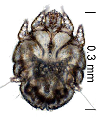
Demodectic mange is caused by the demodex mite, Demodex canis, which lives in normal skin and is present in small numbers in all dogs.  Humans have their own demodex mites (D. folliculorum, D. brevis) and the dog demodex mite never affects humans.

The entire life cycle of the demodex mite is spent on the skin. It lives in hair follicles and feeds on cells, serum and epidermal debris.

Transmission of mites from bitch to puppies occurs during nursing in the first 2 days of life.

Occasionally a puppy’s immature immune system allows the mites to multiply abnormally and mild localised skin disease results.  A small proportion of affected pups have a more depressed immune system and generalised demedicosis develops.

The signs of localised demodicosis include patchy hair loss and mild scaliness on the face.  Sometimes the skin is mildly red but in the early stages it is usually not itchy. Most cases occur before 6 months of age and heal spontaneously.

Up to 10% of affected dogs progress to the generalised form. It usually starts during puppyhood. With generalised demidicosis we see hair loss, inflammation and enlarged glands. Secondary bacterial infections cause scratching, redness, swelling and crusting of the skin.

Dogs with chronic generalised demodicosis have depressed immune T-cell responses. Eradication of the mites results in restoration of the T-cell function.

Breeds predisposed to generalised demodicosis include the Old English Sheep dog, Afghan Hound, Collie, German Shepherd, Staffordshire and Pit bull terrier, Doberman, Dalmatians, Great Dane, English Bulldogs, Boston terriers, Dachshunds, Chihuahua, Boxers, pugs, Sharpeis, Beagles and Pointers. Pure bred dogs have a much higher incidence than mongrels. There is evidence that it is an inherited problem.

To diagnose demodectic mange we scrape affected areas with the side of a scalpel blade and check for mites under the microscope.

We usually do not treat mild localised demidicosis as 90% of cases will clear spontaneously.

If the hair loss spreads and the skin becomes red, itchy or crusty then we treat the pup with a miticide. Regular skin scraping monitors the progress of eradication.  Treatment is continued for at least a month after a negative skin-scraping.

Secondary bacterial infections are treated with antibiotics and antiseptic shampoos as they arise.

Flea control

Control fleas ON the dog:

  • Capstar or Comfortis tablets kill fleas and paralyse flea mouthparts preventing deposition of saliva.
  • Monthly applications of Advantage, Advocate, Advantix, Frontline, Revolution, or Comfortis throughout the year keep flea numbers as low as possible.
  • Flea powders, sprays, and shampoos kill the fleas present on your dog at the time of application but have little residual effect.

Control fleas IN the dog’s environment:

  • Professional fumigation of the house and yard will control egg, larval and pupal stages of the flea life cycle most effectively.
  • If you spray or flea bomb yourself repeat the application in 3-4 weeks to catch the next hatching of pupae.
  • Some products also contain growth regulators that prevent flea maturation.
  • Wash bedding in hot water and dry in the sun.
  • Vacuum carpets and furniture often.
  • Rake up leaves in shady places and provide outside kennels or hidey holes off the ground.
  • Flea larvae love dust so provide working dogs with concrete runs.

Do not be too quick to blame kennels for fleas on your dog. When your pets are gone pupae remain in their cocoon because the house is quiet and empty. When they return the vibrations, heat and light stimulate the emergence of adult fleas. They jump on dogs, cats, and even people looking for a blood meal.

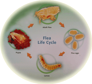
Effective flea control depends on knowing the flea’s life cycle.

The flea life cycle

Adult fleas are only 5% of the entire flea population. The eggs they lay on the dog fall off into the dog’s environment. Flea eggs are pearly white and too small to see without magnification. They hatch into larvae in 1 to 10 days depending on how humid and hot it is.

Flea larvae eat organic debris and adult flea faeces. They avoid direct sunlight and burrow into carpet fibres, grass, branches, leaves, or soil.

Dry conditions kill larvae. Outdoor larval development occurs where the ground is shaded and moist and where flea-infested pets spend a significant amount of time. Indoors, larvae survive best in the carpet or in cracks in the floor.

After 5 to 11 days larvae pupate. While in the cocoon pupae are resistant to insecticides and continue to emerge for 3 months despite insecticide application.

Pupae emerge as adult fleas in another 5-10 days if stimulated by the vibration of passing pets, physical pressure, carbon dioxide, or heat.

When the adult flea emerges it moves towards body heat, movement, and exhaled carbon dioxide. Following the first blood meal, female fleas begin egg production within 48 hours and continue for 100 days.

This entire life cycle (adult flea – egg – larva – pupa – adult) takes 7 – 21 days depending on temperature and humidity conditions.

Flea control depends on knowing this life cycle.

Flea allergy

What is flea allergy?

Some dogs are allergic to fleas and react with non-stop scratching and biting. Normal dogs are only mildly irritated by fleas. The flea allergic dog is allergic to flea saliva. Just one flea bite causes intense and long lasting itchiness.

The dog chews, licks, or scratches over the rump, on the belly and down the legs. This causes hair loss and damage to the skin. Breaks in the skin allow bacteria in and infection to develop.

What is the proper treatment?

We must eliminate all fleas on the dog and in the environment.

Capstar or Comfortis kill the fleas and paralyse flea mouthparts preventing deposition of saliva.

Every month treat your dog with Advantage, Advocate, Advantix, Frontline, Revolution, or Comfortis to keep flea numbers as low as possible.

Fleas spend most of their life cycle off the dog as larvae and pupae in bedding, carpet, dirt and leaves. Wash bedding in hot water and dry in the sun.  Vacuum carpets and furniture often. Rake up leaves in shady places and provide outside kennels or hidey holes off the ground.  Call a professional to fumigate your house.

Flea control depends on knowing the life cycle of fleas.

Cortisone products like prednisolone block the allergic reaction and give relief from the intense itching.

If your dog develops a bacterial infection in the skin, your vet will prescribe antibiotics.

Sarcoptic mange or scabies

Itchy dog?

Sarcoptic mange causes severe itching.  Dogs chew and scratch their skin constantly.  They lose hair and the skin is thick and red, especially on the ears, legs and belly.

What causes sarcoptic mange?

It is caused by a mite that lives on the skin of any age dog.  It’s also called fox mange. Many dogs catch it from foxes.

Is it contagious?

Sarcoptic mange is highly contagious to other dogs. Humans in close contact with their dog  can catch it, too.  Although the mites are not able to complete their life cycle on humans, they cause a rash and severe itching before they finally die.  Contact your family doctor or pharmacist for advice on treatment.

How is sarcoptic mange diagnosed?

Diagnosis is made by scraping the skin with the side of a scalpel blade and examining it under the microscope.  Despite severe itchiness there are often only a small number of mites present on the dog.  If all skin scrapings are negative and we are still suspicious we do a treatment trial.

How is it treated?

The spot on treatment Revolution applied weekly for 3 weeks is easy and effective.  Sometimes we inject or drench with ivermectin weekly. Ivermectin is not licensed for use in dogs and is dangerous for collie breeds.

All dogs in contact with the affected dog should be treated.

Discard the dog’s bedding before treatment.

If any member of the family develops an itchy skin rash, please tell your doctor that you have been exposed to a dog with sarcoptic mange, also known as scabies.

Mast Cell Tumours

Mast Cell Tumours are aggressive skin cancers and common in dogs. They can look like anything: a patch of dry skin, a raised red itchy area, a wart or a lump.

Boxers and Staffordshire Bull Terriers have a higher than average risk of Mast Cell Tumours.  Other breeds affected include the Bull Terrier, English Bulldog and Sharpei but we see Mast Cell Tumours in all breeds.

Mast Cell Tumours release chemicals randomly, causing local redness, itch and swelling that comes and goes.

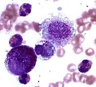
To diagnose a Mast Cell Tumour we take a sample of cells, stain them and examine them under the microscope.  Most Mast Cell Tumours contain characteristic granules that are easily recognised.  If we have any doubts we recommend removal anyway.

The lesion is removed with a wide excision because Mast Cell Tumours send out long microscopic fingers into the surrounding tissue.  It is sent to the pathologist who confirms the diagnosis, makes sure we have removed it all and grades it.

The grade of the tumour reflects how malignant the tumour is.  A low grade tumour is unlikely to have spread and, as long as the entire tumour has been removed, is unlikely to recur.

About 25% of all Mast Cell Tumours are higher grade.  They invade locally and can spread.  Without supplementary chemotherapy, the mean survival time is 18 weeks.

Chemotherapy is effective and median survival times are good.

Itchy Dogs

Many itchy dogs have the doggy equivalent of hayfever, which is called atopy.  A lot of dogs are allergic to flea saliva but only a few are allergic to food.

Common causes, or allergens, include pollen, grasses and dust mites.

Atopic dogs also have a poor skin barrier that allows the allergens to penetrate the skin and provoke the allergic reaction.

How does atopy affect my dog?

Dogs with atopy constantly scratch, bite, rub or chew themselves.  Itchy dogs don’t always scratch. They may rub their faces, flap their ears, or lick and chew their feet.

Some dogs scratch continuously but don’t damage their skin, whilst others quickly rub themselves raw. Recurrent ear infections are common.

Some dogs show signs of atopy as young as 3 months old, but usually it first occurs between 1 and 3 years of age. Initially most dogs will only be itchy during certain seasons, usually spring and early summer. As your dog ages, these itchy periods become longer.

As atopic dogs age, they become allergic to more things. Each dog has an allergenic load he can tolerate before he starts to itch. Exposure to one extra pollen or flea bite pushes him over the threshold into scratching or rubbing.

Diagnosis depends on the information you supply and examination of the skin. Tests such as skin scrapings and cytology rule out mites and secondary infections.

We may also rule out flea and food allergy by eliminating fleas and starting a dietary trial.

Skin testing by a specialist dermatologist will identify the allergens.

How to we treat atopy?

It is rare to cure atopy. We control it with a combination of strategies:

Improving the skin barrier:

  • Use shampoos without sulphates
  • Add essential fatty acids like Nutricoat, Megaderm, and fish oil to the diet
  • Apply sphingosines found in Nutriderm shampoo and conditioner,
  • Minimise bathing as much as possible

Avoiding allergens:

  • Vacuum carpeted areas frequently and minimise bedding to reduce dust mites
  • Keep your pet inside on windy days or when mowing

Treatments:

  • Cortisone offers immediate relief but can have serious side-effects if used long term
  • Atopica has fewer side-effects than cortisone and is often very effective but can be expensive in large dogs in the initial stages
  • Hyposensitization with repeated small injections of low doses of a combination of allergens chosen by a dermatologist on the basis of skin tests. These suppress the allergic reaction successfully in approximately 70% of atopic pets. Relief is not immediate and they usually require injections at least monthly for the rest of their lives. However in responsive pets hyposensitisation minimises the use of other medication.
  • Antihistamines such as Claratyne, Fexotabs, Claramax or Zyrtec are helpful in some cases but are rarely as effective as in humans.
  • Shampoos containing aloe vera and oatmeal may ease itchy skin

Skin allergies can be challenging to control and require a systematic approach to diagnose and treatment.  With commitment and care, most pets can enjoy a comfortable life despite ongoing exposure to environmental allergens.