Category Archives: Dogs

Cushing’s disease

In dogs with Cushing’s disease the adrenal glands overproduce some of the body’s regulators, particularly cortisol.

What are the signs of Cushing’s disease?
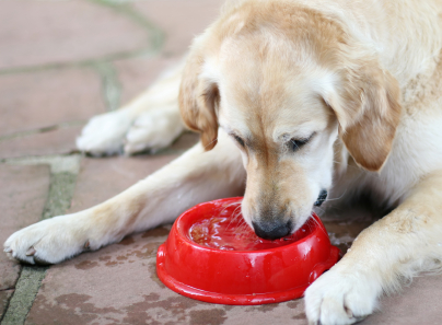
The most common signs of Cushing’s disease are marked increases in appetite, water consumption and urination. Lethargy, panting and a poor hair coat are also common. We often see a pot-bellied or bloated abdomen due to increased fat within the abdominal organs and thinning of the muscular abdominal wall.

What causes Cushing’s disease?

The three major causes of Cushing’s Disease:

  1. A tumour of the pituitary gland, that stimulates the adrenal glands to produce excessive  amounts of cortisol.
  2. Excessive administration of synthetic cortisones cortisones such as prednisolone, triamcinolone or dexamethasone may cause Iatrogenic Cushing’s disease.
  3. An adrenal gland tumour is an uncommon cause of Cushing’s Disease.

If we suspect Cushing’s Disease we run a blood test to check your dog’s general health. An enzyme called Alkaline Phosphatase (ALKP) is often high in dogs with Cushing’s disease. A Low Dose Dexamethasone test (LDDT) confirms or denies Cushing’s disease.

To determine which type of Cushing’s disease your pet has, we ultrasound the adrenal glands and/or do an endogenous ACTH blood test.

What are the treatment options?

Pituitary Tumour: This is the most common cause of Cushing’s disease. There are two treatment options for it. Trilostane is our drug of choice. A daily capsule of Trilostane reduces the production of cortisone and another important hormone, aldosterone.  We monitor your dog’s response to Trilostane with a test called the ACTH stimulation test. Too little Trilostane won’t reduce appetite or water consumption but too much will cause illness.

If your dog has liver or kidney disease we may suggest treatment with Mitotane (also known as Lysodren). This drug destroys part of the adrenal gland. Careful monitoring and good communication with your vet is necessary during the initial intensive treatment to achieve good results and avoid life-threatening adrenal damage.

Although the pituitary tumour remains present and continues to stimulate the adrenal gland if the tumour is small successful control for many years in most dogs is possible.  If the tumour is large, it may invade surrounding brain tissue and cause other signs, but this is rare.

Iatrogenic Cushing’s Disease: To treat this type of Cushing’s disease we must stop the synthetic cortisone in a very controlled way. If we stop intensive cortisone treatment abruptly your dog may lose his appetite, vomit, develop diarrhea and collapse. The suppressed adrenal gland takes a while to regain normal production of cortisol.

Treatment of an Adrenal Tumour:

Adrenal tumours tend to invade surrounding tissue but if we can surgically remove it all and it is not malignant your dog will regain normal health. Otherwise we treat adrenal tumours with Trilostane also.

Boarding your pet

Holiday plans are not complete without accommodation arrangements for your pets, too. Many holiday accommodations are now pet friendly but most people have to leave their pets behind. Ideally, they should stay in their own home and yard with a friend, relative or house-sitter looking after them.

Many pets spend happy holidays at boarding facilities and even look forward to their own break from home routines. If possible visit the cattery or kennels beforehand, inspect the accommodation and meet the staff.

All commercial boarding establishments are licensed by the local authority but standards vary. Seek out recommendations from friends, neighbours or your veterinary surgeon. Some people like to trial board their dogs for a weekend or a few days and see how they settle.

Check that your pets’ vaccinations are up-to-date well before the holiday. Kennels insist upon current cover for distemper, hepatitis, parvovirus and kennel cough for dogs and enteritis, calicivirus and herpesvirus for cats.

A single intra-nasal kennel cough immunization at least 72 hours before boarding covers dogs previously unvaccinated for kennel cough for 12 months. Other vaccinations take at least ten days to take effect.

Itchy bottom?

Dogs scoot their bottoms along the ground when they have anal gland problems, tapeworms or allergies.

If you catch your dog rubbing along on the carpet, worm him with a good quality wormer such as Drontal or Milbemax that covers all worms, especially tapeworm.

If he still scoots or seems off colour then bring him in to the surgery. Many small dogs suffer from anal sac problems. The gland fills up with material too thick to empty through the small ducts near the anus. Usually we just express them and all is well.

Sometimes the material gets infected and the glands become swollen and painful.  Your dog might have trouble defecating or lick the area a lot. The glands may break through the skin and discharge foul smelling fluid. At the surgery we clip and clean them as well as starting antibiotics and pain relief. If your dog has repeated anal gland infections we recommend surgery to remove them.

In spring and summer many allergic dogs rub, lick and scratch all over including their bottoms. Some dogs allergic to food proteins also rub their bottoms on the ground. Treatment for the allergies usually stops the rubbing and licking.


Inflammatory bowel disease

Inflammatory Bowel Disease (IBD) is a chronic disease of the gut, or gastrointestinal tract. Most affected dogs have a history of recurrent or chronic vomiting and/or diarrhoea. They may lose weight but are otherwise normal. Most eat well or have an increased appetite, and appear normal.

Cause

Inflammatory cells invade the lining of the bowel causing thickening of the bowel. This interferes with digestion and absorption of nutrients and with normal movement of the bowel.

When the cells invade the stomach walls or upper small intestine vomiting develops. Infiltration of the small intestine causes watery diarrhoea and weight loss. Infiltration of the large intestine causes diarrhoea coated with mucus and fresh blood. The entire gastrointestinal tract is sometimes affected.

Something in the diet triggers the disease in some dogs. Bacterial proteins are involved in other cases. In most cases we cannot find any underlying cause.

Diagnosis

IBD is diagnosed by ruling out other diseases. Blood tests rule out liver or kidney disease, pancreatic insufficiency and diabetes. Faecal tests rule out parasites like whipworms or giardia.

X-rays or ultrasound rule out some tumours or cancers in the abdomen.

If these tests are all clear we trial special low allergy, low residue or high fibre diets, depending on the part of the bowel most involved.  If bacterial overgrowth is present we trial various antibiotics until we see improvement.

If there is no improvement we biopsy the bowel and a pathologist identifies the type of cells involved, assesses the severity of the inflammation and checks for cancer cells.

Treatment

If any diet has improved the condition we continue it.

Medication controls (not cures) the problem. We trial drugs like metronidazole, prednisolone and azathioprine until we find what suppresses the signs best. Most dogs stay on the appropriate drugs and diet for life.

Hypothyroidism

The thyroid gland regulates the body’s metabolic rate. When it slows down the whole body slows down.

Hypothyroidism is a deficiency in production of the thyroid hormone. This deficiency is caused by immune-mediated destruction of the thyroid gland, most commonly, or by natural atrophy of the gland.

Signs of hypothyroidism include:

  • Weight gain
  • Lethargy
  • Cold intolerance
  • Dry coat and excessive shedding of hair
  • Very thin coat
  • Increased pigmentation of the skin
  • Failure to re-grow hair after clipping or shaving
  • A tragic expression because of thickening of the facial skin
  • Abnormal nerve function exhibited as non-painful lameness, dragging of the feet, lack of co-ordination and a head tilt
  • Loss of libido and infertility in intact males
  • Lack of heat periods, infertility, and abortion in females
  • Spots on the eyes
  • Dry eye because of inadequate tear production.
  • Anaemia

The thyroid glands are located in the neck either side of the trachea or windpipe. They are controlled by the body’s master gland, the pituitary gland, located at the base of the brain.


To confirm hypothyroidism we test blood to see if T4, the main thyroid hormone, is low. Unfortunately, dogs sick for some other reason or taking certain drugs like seizure medications, may have low levels too. Confirmation of hypothyroidism sometimes requires an additional test.

Hypothyroidism is treated daily with tablets containing a thyroid replacement hormone, thyroxine, for the rest of the dog’s life.

We check T4 levels again after a month of treatment to make sure we are giving the right dose. We test 4-6 hours after the morning dose. T4 levels are then checked every 6 months and the dose adjusted if necessary.

Overdosing produces signs of hyperthyroidism including hyperactivity, lack of sleep, weight loss, panting, nervousness, aggressive behaviour and an increase in water consumption. If any of these occur, please let us know immediately.

Demodectic mange

Demodectic mange is caused by the demodex mite, Demodex canis, which lives in normal skin and is present in small numbers in all dogs.  Humans have their own demodex mites (D. folliculorum, D. brevis) and the dog demodex mite never affects humans.

The entire life cycle of the demodex mite is spent on the skin. It lives in hair follicles and feeds on cells, serum and epidermal debris.

Transmission of mites from bitch to puppies occurs during nursing in the first 2 days of life.

Occasionally a puppy’s immature immune system allows the mites to multiply abnormally and mild localised skin disease results.  A small proportion of affected pups have a more depressed immune system and generalised demedicosis develops.

The signs of localised demodicosis include patchy hair loss and mild scaliness on the face.  Sometimes the skin is mildly red but in the early stages it is usually not itchy. Most cases occur before 6 months of age and heal spontaneously.

Up to 10% of affected dogs progress to the generalised form. It usually starts during puppyhood. With generalised demidicosis we see hair loss, inflammation and enlarged glands. Secondary bacterial infections cause scratching, redness, swelling and crusting of the skin.

Dogs with chronic generalised demodicosis have depressed immune T-cell responses. Eradication of the mites results in restoration of the T-cell function.

Breeds predisposed to generalised demodicosis include the Old English Sheep dog, Afghan Hound, Collie, German Shepherd, Staffordshire and Pit bull terrier, Doberman, Dalmatians, Great Dane, English Bulldogs, Boston terriers, Dachshunds, Chihuahua, Boxers, pugs, Sharpeis, Beagles and Pointers. Pure bred dogs have a much higher incidence than mongrels. There is evidence that it is an inherited problem.

To diagnose demodectic mange we scrape affected areas with the side of a scalpel blade and check for mites under the microscope.

We usually do not treat mild localised demidicosis as 90% of cases will clear spontaneously.

If the hair loss spreads and the skin becomes red, itchy or crusty then we treat the pup with a miticide. Regular skin scraping monitors the progress of eradication.  Treatment is continued for at least a month after a negative skin-scraping.

Secondary bacterial infections are treated with antibiotics and antiseptic shampoos as they arise.

Haemorrhagic gastroenteritis

Dogs affected with haemorrhagic gastroenteritis (HGE) vomit and pass diarrhoea with blood in it.  It develops rapidly and sometimes owners just find their dog collapsed.

HGE is more common in small breeds of dogs. A very high red blood cell concentration, measured as the packed cell volume, PCV, or haematocrit is characteristic of HGE. Most normal dogs have hæmatocrits of 37-55%. Dogs with HGE have hæmatocrits over 60%.

We don’t know the exact cause of HGE.

We rule out other causes of vomiting and bloody diarrhoea while we start emergency treatment for HGE.  These causes include stomach ulcers, trauma, worms, other gut parasites, gastrointestinal tumours, intestinal obstruction, foreign bodies, parvovirus, and coagulation disorders.

Dogs with HGE are very ill and if left untreated die. In most cases, it runs its course in a few days with intravenous fluid therapy to combat the dehydration and shock. We give other treatments like antibiotics, anti-ulcer medication and corticosteroids as indicated.

If intravenous fluid therapy is not given the haematocrit continues to rise and the blood becomes so thick that it can hardly flow. A clotting disorder called disseminated intravascular coagulation develops which is often irreversible and fatal.

Corneal ulcers

The cornea is the transparent front part of the eyeball that admits light to the eye.

A corneal ulcer is a hole in the top layer of the cornea. The most common cause of corneal ulcers is trauma. Grass seeds, a cat scratch, shampoo and sticks cause most of the corneal ulcers we see at Hall Vet Surgery.

Dry eye develops because of abnormal tear formation in older dogs and is also a common cause of corneal ulcers.  Some diseases that affect the whole body like diabetes mellitus, Cushings disease, and hypothyroidism sometimes predispose a dog to corneal ulcers, too.

A corneal ulcer is very painful.   The affected dog rubs the eye and squeezes it tightly closed. The eye looks red and watery.

Superficial corneal abrasions are difficult to see. A drop of fluorescein stains the area of ulceration and shows it up clearly under a special blue light.

A superficial corneal abrasion generally heals within 5 days. Antibiotic eye drops or ointment prevent bacterial infections.  Atropine eye drops or ointments relieve spasm and pain.

If the corneal ulcer is deep or slow healing or a descemetocele has formed we protect the eye and promote healing with a surgical overlay of conjunctiva, the third eyelid or the upper and lower eyelids.

Atropine relieves the pain from the ulcer but dilates the pupil making the dog sensitive to light. Do not be alarmed if the pupil stays dilated for several days after the last dose.

Atropine travels down the tear ducts to the mouth and because it tastes bitter causes drooling and pawing at the mouth in some dogs.

When the fluorescein stain test is negative your vet will tell you to discontinue the treatment. This is usually after at least 5 days of treatment.

The normal cornea has no blood vessels in it. When it is ulcerated blood vessels grow in from the white part of the eye, the sclera, to heal it and may obstruct vision. If they don’t retreat once the ulcer is healed we clear them with cortisone drops or ointment.

Collapsing tracheas

Collapsing tracheas are common in small dogs like Chihuahuas, Pomeranians, Shih Tzus, Lhasa Apsos, Toy Poodles, and Yorkshire Terriers.

The trachea, also known as the windpipe, connects the throat to the lungs. The normal trachea is tubular and maintains its shape with a series of “C” shaped rings made of cartilage.

When a dog with a collapsing trachea breathes in, the cartilage rings flatten from top to the bottom. This makes it difficult for air to enter the lungs and causes a chronic, honking cough.

In affected dogs the cartilage rings lose their stiffness and cannot retain their circular shape.

Coughing is often worse in the daytime. The cough is exacerbated by excitement, drinking water, eating, or pressure on the trachea, particularly from a leash.

We suspect a collapsed trachea in a small dog with a persistent dry and/or honking cough. If light pressure over the trachea causes a cough we recommend a chest X ray to rule out other heart, lung and airway problems. These other conditions are common in small dogs and they often suffer from several of them at a time.

Endoscopy allows us to watch the inside of the trachea during inspiration and expiration for signs of collapse.

We treat a dog with collapsing trachea with drugs to open up and reduce inflammation of the airways.

Antibiotics control infections of collapsed airways, which are common.

Weight loss reduces pressure on the airways in obese dogs. A low fat diet and mild exercise helps them shed the extra kilos.

Avoid exacerbating the cough with excitement and vigorous exercise.

Because medical therapy only treats the cough and does not correct the problem, affected dogs will always have a bit of a cough. Our aim is to minimize the cough, and make them more comfortable and active.

Bladder stones

Bladder stones form from minerals excreted into the urinary bladder. They range in size from grains of sand or tiny pebbles to a large, single stone.

The main signs of bladder stones are blood in the urine (hæmaturia) and straining to urinate (dysuria). Urinary tract infections also cause hæmaturia and dysuria.

Hæmaturia occurs because the stones mechanically irritate the bladder wall and make it bleed.

Dysuria occurs when stones obstruct the passage of urine out of the bladder. Large stones sometimes cause a valve-like obstruction at the neck of the bladder. The dog passes some urine and then despite straining cannot pass more. Small stones flow with the urine and obstruct the urethra, especially of male dogs.

Stones form from a high concentration of crystals in the urine. The crystals form because of abnormalities in the diet or because of disease in the bladder, especially bacterial infection. Sometimes they form because of a fault in body chemistry.

We suspect bladder stones if the bladder is painful or when there are recurrent infections. We can feel some bladder stones through the abdominal wall but most bladder stones are diagnosed with x-rays or ultrasound.

The fastest treatment is surgery to open the abdomen and bladder and pick out the stones.

Alternatively, if the dog has passed a stone in the urine we analyse it and prescribe the specific diet that dissolves that type of stone. Diet is not successful in dissolving some kinds of stone and is not fast enough if the pet is already toxic from a back up of urine in the urinary tract.

Some types of stone can be prevented. Regular tests for urinary tract infections and appropriate antimicrobial treatment prevent stones formed as a result of bacterial infection.

Specially formulated diets help prevent the formation of some other types of stone. We determine the chemical composition of stones removed at surgery and prescribe the best preventative diet for that particular type of stone.