Warning: this blog post contains graphic surgical images
Crookshanks is a lovely cat that came in to see us as she had been losing weight and her owners had noticed blood in her urine. After running blood and urine tests we then performed an abdominal ultrasound.
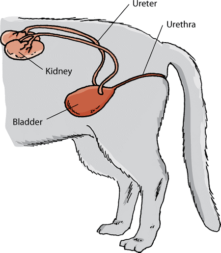
The ultrasound highlighted that Crookshanks had a stone in her ureter – the ureter is the tube through which urine flows from the kidneys to the bladder. The diagram below shows the kidneys, ureters and bladder.
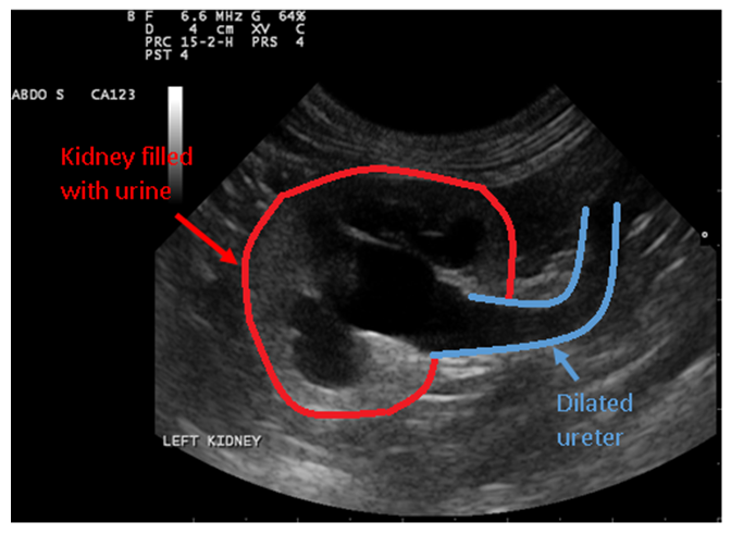
When the ureter becomes blocked the kidney fills with urine and cannot empty. This was also the cause of the blood Crookshanks’ owner noticed in her urine.
On ultrasound a normal kidney is bean shaped, it has a light grey outer shell, and a darker middle area. In the images below the kidney outline is circled in red.
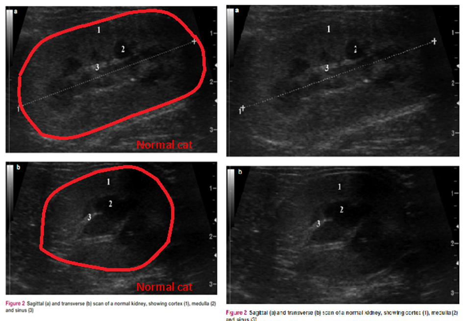
Below are ultrasound images of Crookshanks’ kidney. Again, the outline of the kidney is highlighted in red. The centre of the kidney is black, this is the urine that is building up because her ureter is blocked. Her ureter is widened and full of urine. The ureter is outlined in blue in this image.

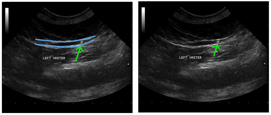
We followed the widened ureter down towards the bladder wih the ultrasound probe. This is when we found the stones that were causing the blockage. The ureter is outlined in blue, and there is a green arrow pointing towards the stones.
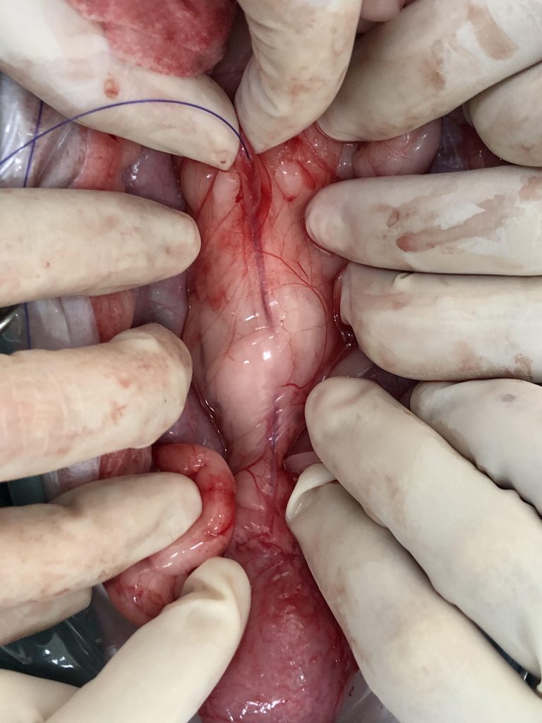
Crookshanks’ went into surgery with Dr Vickie Saye to have the stone removed. The ureter is very tiny, only a few millimeters thick, so it was a very intricate and delicate surgery. You can see in the photo below, the ureter is so small that you can only fit a very fine piece of suture material through it. The photo was taken during Crookshanks’ surgery and the suture material was passed through the ureter to ensure that all stones had been removed and the ureter was blockage free.
Two stones were removed from Crookshanks’ ureters. They were very small only 1-2mm in size, amazing that something so small can cause such big problems.
Here are the two stones surgically removed from Crookshanks’ ureter. There were TINY, about 1mm in size.
Crookshanks required intensive care and monitoring after her surgery. She was in hospital for several days.
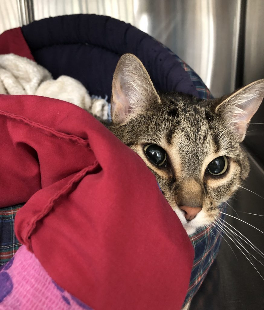
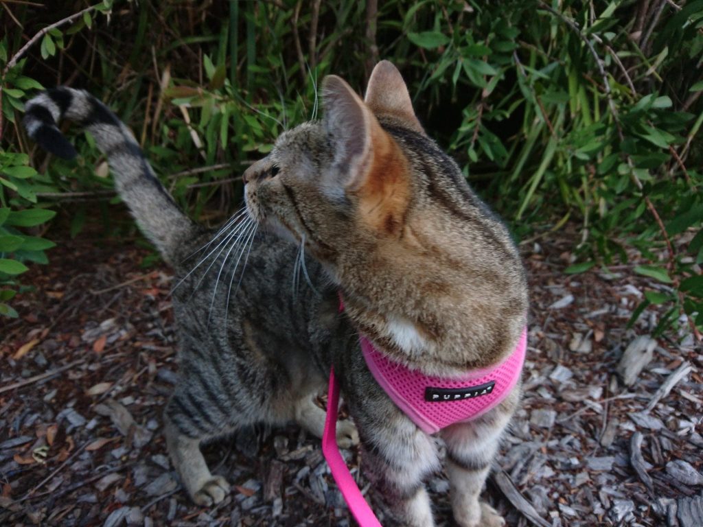
She is now much brighter and happier than she was before the surgery, as she is not in pain anymore. She is even starting to put on weight. She enjoys being able to join her sister McGonagall for walks on lead around the suburb. She will be on a special prescription diet for the rest of her life to help prevent the stones from reoccurring.